This month’s maker is Lia K. Jacobson, MD, MSGM, assistant clinical professor, Division of Pediatric Otolaryngology within the Department of Otolaryngology. We caught up with Dr. Jacobson to see what they made.
Q: What did you make?
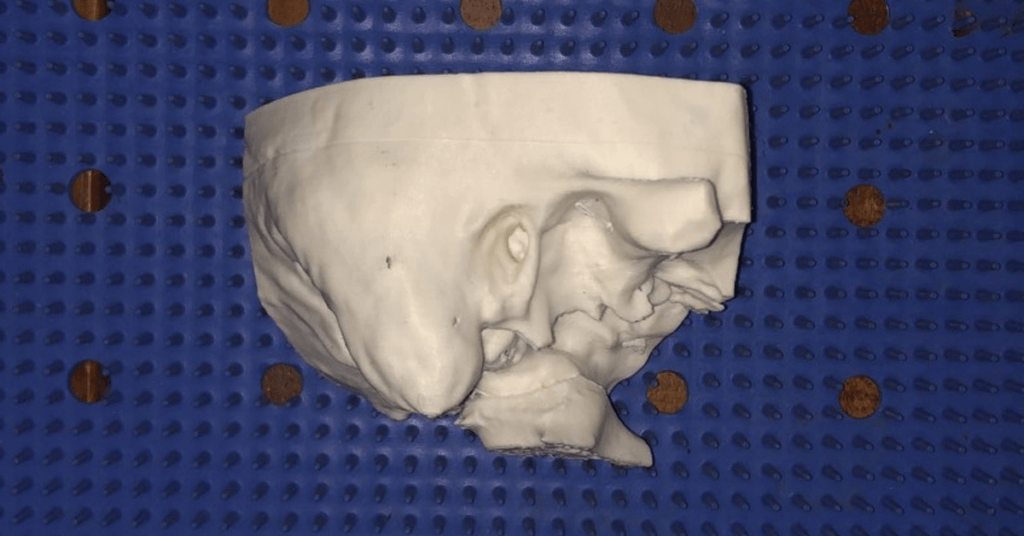
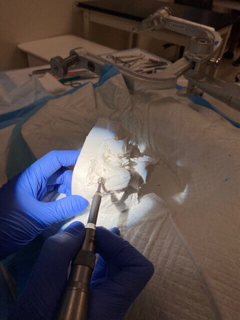
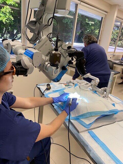
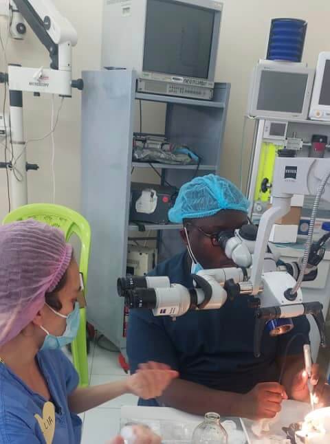
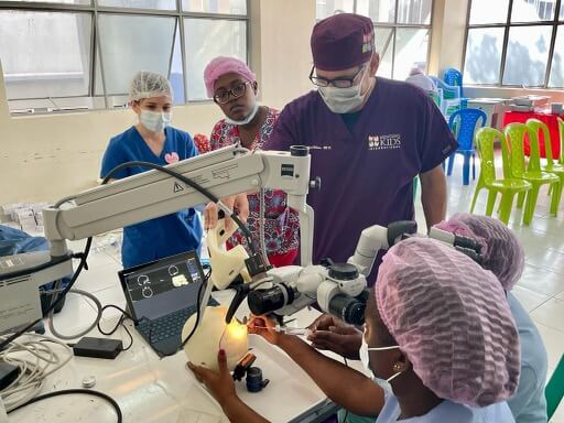
We made anatomically accurate, 3D printed temporal bone models that we used to augment a surgical skills simulation lab for practicing ear surgery techniques. We specifically used these models as a pilot training model for otolaryngologists (ear, nose and throat surgeons) and trainees from low- and middle-income countries. Our goal was to create a high fidelity, affordable and reproducible surgical skills training model that can help teach safe surgical techniques for surgical trainees in resource-limited surgical settings.
Q: Why did you want to make it?
I wanted to pursue this project to leverage UCSF’s fantastic resources to support surgical education worldwide in creative and innovative ways. This project brought together partners in otolaryngology from around the world to supplement surgical training for participants from Tanzania, Ecuador, Uruguay, Ethiopia and Jamaica in a three day temporal bone drilling workshop.
Q: What was your process?
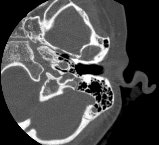
We used CT scan imaging to create a blueprint for our models, and used printing materials that have been validated in previous studies to be similar in feel to bone. This was important as these models were used to teach safe drilling techniques on the temporal bone, the skull bone that house the delicate structures of the middle and inner ear.
Q: What was the hardest part of the process?
I don’t really have an answer for this – was fairly easy once I looped in the right people to help (my awesome resident Abel David with technical aspects of the CT scan template, otologist colleague Jeff Sharon for advice on materials/design, and the Makers Lab!)
Q: What was your favorite part of the process?
This was a great collaboration between the Makers Lab, colleagues at UCSF and colleagues overseas in Tanzania and elsewhere interested in using a low-cost 3D printed model to increase volume and accessibility to practicing safe ear surgery for early career surgeons and trainees in low-resourced surgical settings.
Q: How did this help make you be better at your job?
Working in the field of global surgery often requires getting creative to provide excellent patient care and education with very limited resources. I think having a resource like the Makers Lab to brainstorm and strategize on developing surgical simulation models that can be utilized and transferred to low resource surgical settings is exciting in terms of bringing in fresh ideas to meet complex challenges of working in these settings.
Q: What do you want to make next?
I have some colleagues working on models for challenging pediatric airway situations. I think working with them to use these models in the global health setting would be great.