
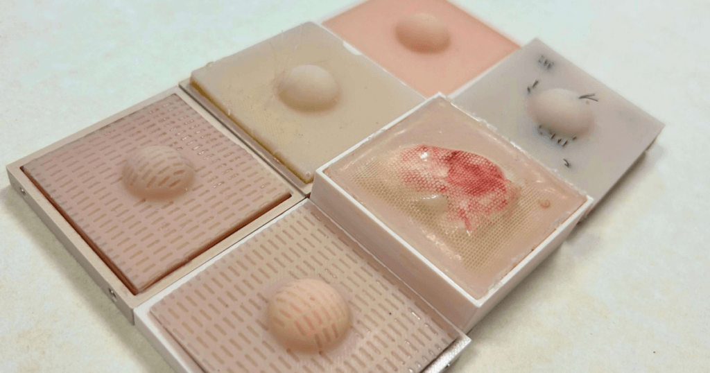
This week’s maker is Alice Tang MD, PhD, a former student at the UCSF Medical Scientist Training Program (MSTP), and now an internal medicine preliminary resident at Kaiser Santa Clara, and a future dermatologist. Let’s take a look at her project:
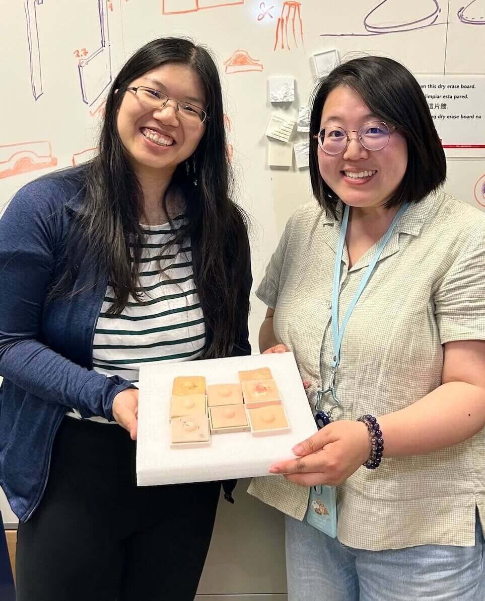
Jenny Tai (right)
Q: What did you make?
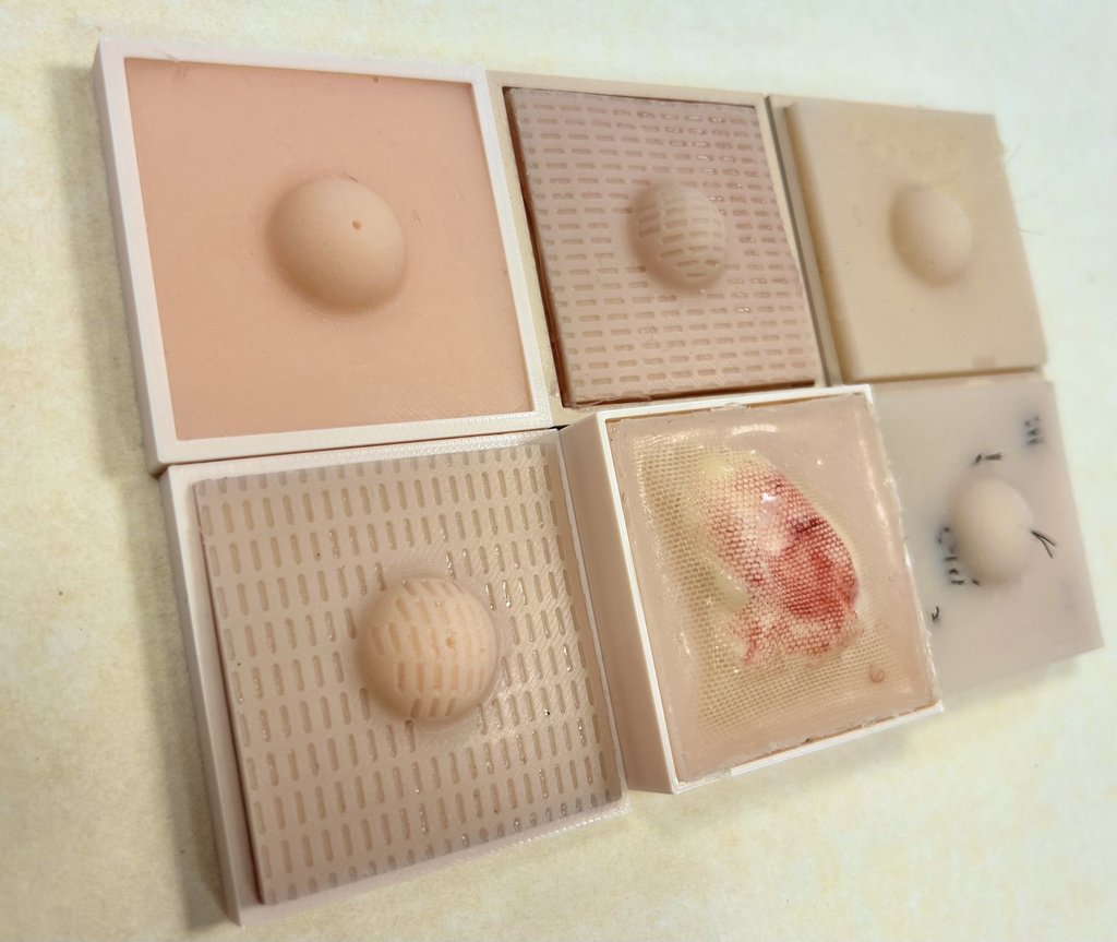
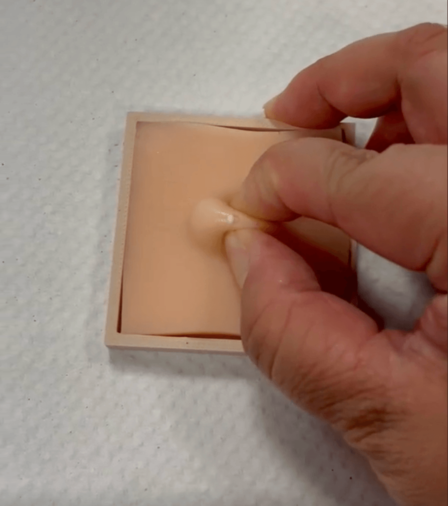
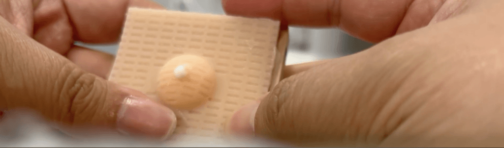
We made tactile 3D kodachromes of various skin conditions. Historically, the term “kodachromes” has been used to describe two-dimensional photos of skin diseases used to teach dermatology. Wax casts like those on display at the Musée des moulages de l’hôpital Saint-Louis are also used to illustrate skin conditions in dermatological education, but those models are not meant to be touched. We have tried to represent skin conditions such as cysts and lipomas with lumps/bumps in our first prototype, while also including visually adjacent conditions like infantile hemangiomas.
Q: Why did you want to make it?
Photographs of skin conditions sometimes do not capture the reality of skin textures or diagnoses that are based on tactile experience. Existing tactile simulations of skin, such as the suture kits we usually get in medical school to practice surgical skills, are not typically representative of real human skin. While learning dermatology, I was missing that 3D tactile element and had to build on my own experiences with patients. I believe more tactile tools can be helpful for medical education, medical students, residents, or any other trainees. Also, since dermatologists are scarce, I believe these tools can help non-dermatologists evaluate skin conditions.
Q: What was your process?
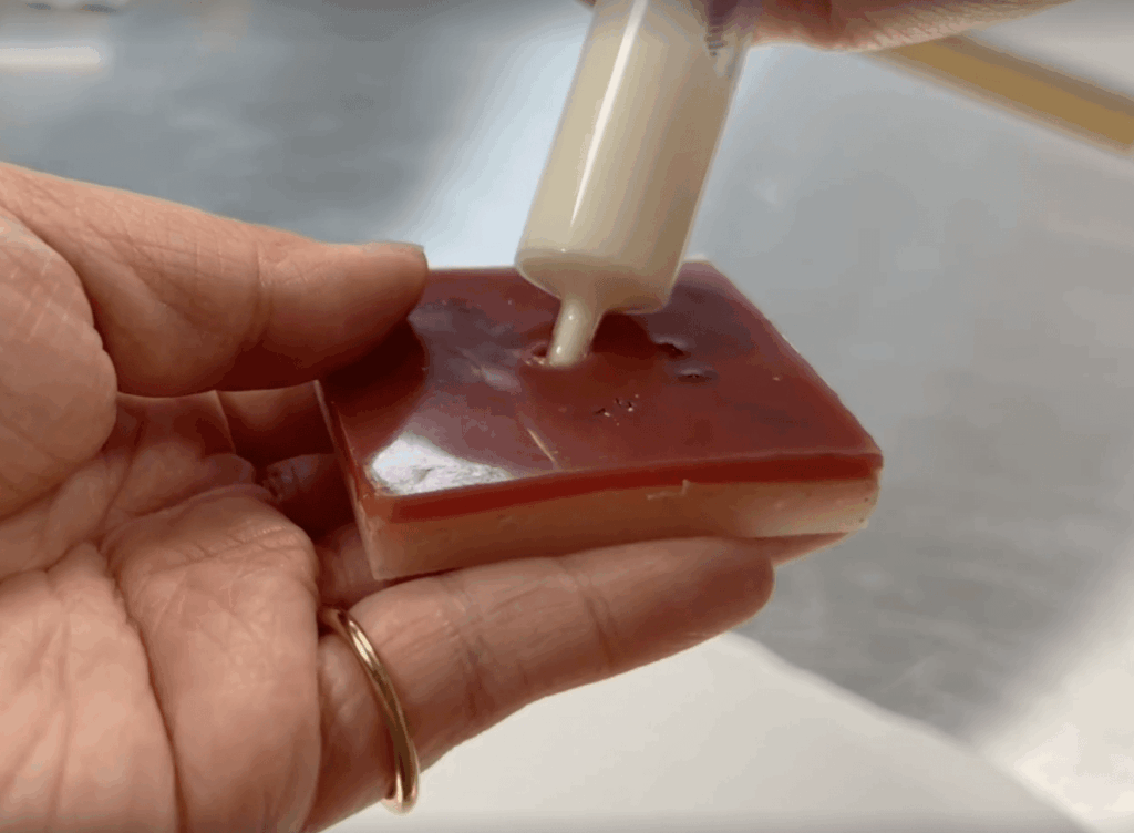
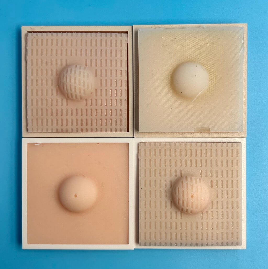
I was inspired by the recent rise of sensory toys, and by my own love of textured touch-and-feel books you usually find for toddlers (which I enjoy as an adult). I was fortunate to meet Jenny Tai, Engineer from the Makers Lab, and we decided to prototype sebaceous cysts and lipomas to start. These could be represented by simple sphere shapes that were hollowed out using 3D modeling software, and small two-part mold trays. Silicones of varying hardnesses can be poured into the spheres to simulate lipomas, while liquids like lotion can be injected into the void space to simulate fluid-filled cysts. Making each skin condition sample a modular square helps to reinforce that kodachrome-esque feel.
Q: What was the hardest part of the process?
I think the hardest part was getting the skin texture right. We looked at existing models, such as suturing kits, as an obvious starting point, but they did not represent skin well. The material is often stretched tight and does not have the looseness of real skin. We also experimented with layering various materials (e.g., silicone bandages) to represent texture differences based on skin depth. Skin often moves, folds, and gives when you press on it, and that pliability contributes to the texture of how skin conditions can behave. Uniform or firm materials may represent lesions, scarring, cancer, or other conditions.
With Jenny’s help and inspiration from prior silicone-based Makers Lab projects, we experimented with various materials and patterns until we felt like we were getting closer to actual skin. We also tried to represent hair and bumpy skin texture using monofilament beading cord sewn into mesh fabric. Optimizing the realism of these models is an ongoing process.
Q: What was your favorite part of the process?
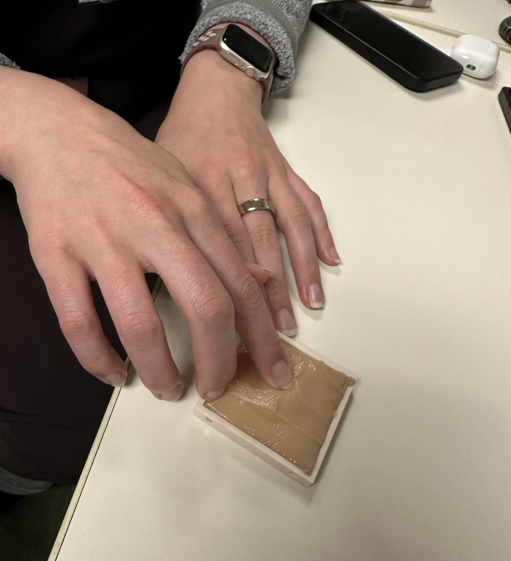
Honestly, watching people’s reactions after our prototype was created! Not only were the “dermachromes” educational, but they also provided great material for debate and intellectually stimulating conversation when I shared them with my medical colleagues. Beyond that, I feel many of my peers take joy in the curiosity and excitement when they touch the models. That has been my favorite part!
Q: How did this help make you a better physician-scientist?
I think this may help me become a better physician because it supports my own education, and helps educate non-dermatologists and possibly patients. I also learned some of the engineering aspects of material selection and of thinking about reproducibility/scalability from the Makers Lab.
I’d love to use these 3D textures to help explore why a texture exists in the first place. For example, I think the models can facilitate understanding of sequelae or chronic health conditions that develop as a consequence of a prior disease, injury, infection, or treatment, such as inflammation, swelling, fibrosis, etc. Understanding involved biomolecules and processes can also help with understanding the possible connection between texture and the causes of disease.
Q: What do you want to make next?
I want to expand to include more skin conditions! I’m thinking about psoriasis, actinic keratosis, ulcers, skin cancers, and more. Stay tuned!
Acknowledgment by Alice Tang
I would like to thank Ernest Y. Lee, MD, PhD, for his mentorship.